
And the Winner Is...
In January, we hosted our very first science art contest! We had three categories contestants could choose from based on their location: Davis/Sacramento, California, or out-of-state (honorable mention only). We received incredible submissions to all categories, and are excited to announce the winners!
Davis/Sacramento:
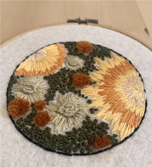
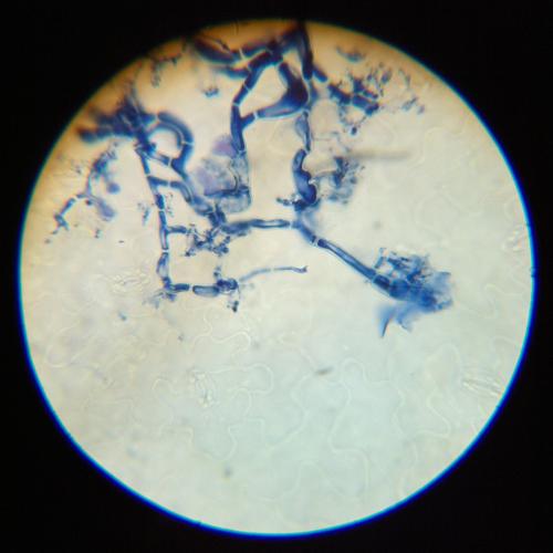
Jayce Taylor won our local Davis/Sacramento category with her embroidery piece of a forgotten petri dish (we’ve all been there!).
What’s the science behind the image?
I'm a chemistry graduate student in the Atsumi lab here at UCD. Our research is in the field of metabolic engineering, with the primary goal of genetically engineering microbes to produce fuels, sugars, pharmaceuticals, and other chemicals from sustainable sources. An important tool in the field of metabolic engineering is antibiotics and antibiotic resistance. When genetically engineering a microbe, scientists will often introduce a gene for antibiotic resistance along with the main gene of interest. When grown on a petri dish containing the antibiotic, only microbes containing the antibiotic resistance and gene of interest will grow. This allows for us to easily select microbial colonies that have our gene of interest or gene edit! Unfortunately antibiotics don't stop fungal growth, so if plates become contaminated, fungi will start to grow and take over. This piece was inspired by a plate forgotten in the back of our lab fridge. Although the fungus was cute and fuzzy, it was too risky to keep around as a "pet" and was subsequently autoclaved.
How important is art in your science journey?
Growing up I always wanted to become either a scientist or an artist, and even though I ended up pursuing science as a career, I continue to work on art in my free time. People often think of science and art as opposite disciplines, but they have a lot in common. Failure is a big theme between the two, and both science and art have taught me how to fail and creatively overcome failure by trying different techniques or methods. I particularly enjoy art projects that I can use, such as ceramics, sewing, and various fiber arts.
California:
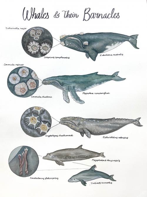
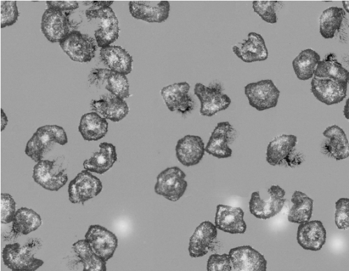
Jenn Cossaboon won our California category with her illustration of whales and their barnacles.
What’s the science behind the image?
About a month ago, I was admiring a photo of a right whale (I am a sucker for charismatic megafauna) and was completely floored to learn that the barnacles found on whales belong to species that have evolved to reproduce, develop, and survive only on whale skin! Whales are actually migrating biomes that host many clingy crustaceans including whale barnacles (which belong to their own subfamily, Coronulidae). Because they cannot leave, these barnacle tenants have synchronized their daily behavior, body structure, and entire life cycle to be compatible with their cetacean hosts. Each species has a unique appearance and preference for certain whale species, which I focused on for the illustration (and took a surprising amount of time to research!). If you're excited about these charismatic microfauna and want to learn more - I wrote a short article on whale barnacles for this week's Creature Feature blog hosted by the awesome UCD Animal Behavior grad students.
How important is art in your science journey?
Art has been a very important part of my science journey both for continuing to learn about/honor wildlife species and to decompress from the pressures of academic life. I took a marine science illustration and watercolor class at UC Santa Cruz during undergrad and it ended up being truly life changing for managing burnout. I am also a highly visual learner, so drawing out anatomy and physiology concepts has been instrumental to my animal medicine and research training - which I hope to continue incorporating scientific illustration into in the future!
Out-of-state:
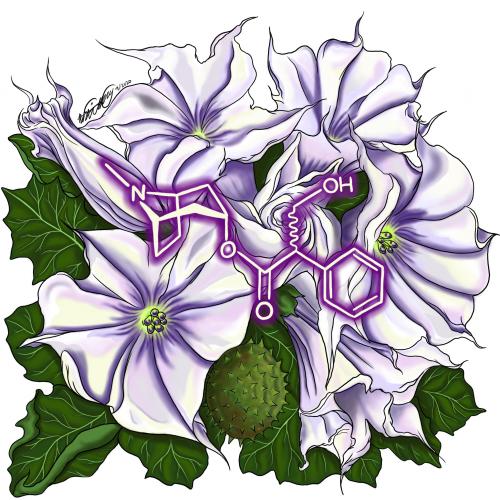
Brandi A. Bundy won our out-of-state competition with her illustration of Jimsonweed with the molecule atropine superimposed on top of the flowers.
What’s the science behind the image?
This globally recognized molecule shares a connection with a plethora of living organisms and this piece is a way to celebrate that. Datura is a member of the nightshade family which is notorious for being deadly. It is fun to superimpose a widely useful alkaloid on a plant commonly associated with death. As an undergraduate I had always been fascinated by alkaloids and toxins. I found that connecting my love for art and chemistry was the best way to satisfy this curiosity.
How is art important for you in your science journey?
Art is important because it has been a timeless medium of communication. There can often be a disconnect or apprehensiveness when it comes to science and I believe art is a great way to bridge the gap for a general audience. Science art is a way to show individuals that science is not something to be feared, hated, or avoided but embraced. My hope is that viewers gain a new perspective of the world around them on a more intimate scale with a growing appreciation for science.
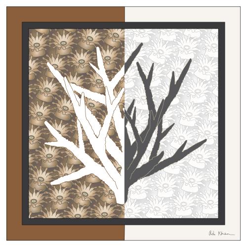
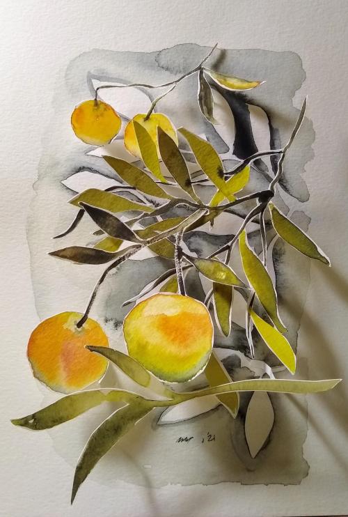
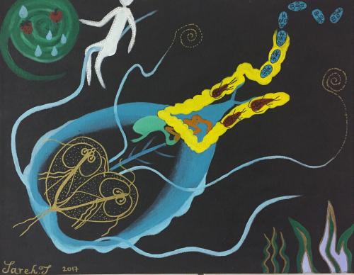
If you missed out on voting, check out all the submissions below.
Davis/Sacramento:
California:
Out-of-state:
Sydney Wyatt is a PhD student at the University of California in Davis. For more content from the UC Davis science communication group "Science Says", follow us on Twitter @SciSays.
Comments